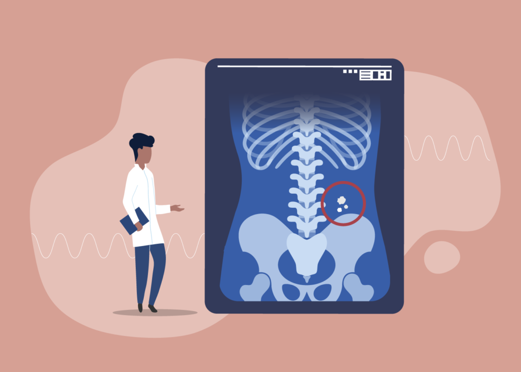
Diagnosed with a kidney stone, but confused about why it didn’t show up on your initial KUB (kidney, ureter and bladder) X-ray? It all boils down to its radio-opacity! A small percentage of the kidney stones go undetected, as radiopaque stones are easier to detect on X-ray than radiolucent stones.
How does radio-opacity affect my X-rays?
Radio-opacity is determined by a structure’s ability to absorb radiation, which affects its visibility on X-rays.
Radiopaque structures, including kidney stones, are opaque and will appear as white areas on X-rays. Your bones, for example, are radiopaque. That’s why you can see them clearly and easily on any X-rays.
Radiolucent structures, in contrast, can absorb radiation and will appear grey or black. This makes it harder for physicians to distinguish your radiolucent kidney stone from fat or soft tissue.
Does my stone type affect its visibility on X-rays?
Yes!
Calcium stones, such as calcium oxalate and calcium phosphate stones, are radiopaque, and can be easily detected through radiography1.
As calcium stones are the most common form of kidney stones, majority of patients can rest assured that their stone is likely to turn up on their X-ray.
Cystine and struvite stones can have variable visibility on X-rays, while pure uric acid and indanivir stones are radiolucent1, which means that they cannot be detected on X-rays at all! Uric acid stones make up approximately 10% of all kidney stones, and are most commonly found in patients who have insulin resistance or a higher BMI. Indanivir stones are caused by indanivir sulfate, which is used in the treatment of HIV (human immunodeficiency virus).
If you know the Hounsfield unit (HU) of your stone, research suggests that that the minimum value of density at which the stone can be detected in the radiographic examination is 498.5 HU2.
What happens if I have a radiolucent stone?
Don’t fret! X-rays are not the only imaging technique used in diagnosing kidney stones.
Based on guidelines from the American Urological Association, a non-contrast CT scan will be administered, possibly in conjunction with the X-ray3.
CT scans have the highest sensitivity for detecting kidney stones, among the various imaging techniques available4.
Curious to learn more about imaging tests for your kidney stones?
Download your free copy of our Survivor’s Guide: Volume 2 here.
Sources:
- Parmar, M. S. (2004, June 12). Kidney stones. BMJ (Clinical research ed.). Retrieved June 2, 2022, from https://www.ncbi.nlm.nih.gov/pmc/articles/PMC421787/
- Chua, M. E., Gatchalian, G. T., Corsino, M. V., & Reyes, B. B. (2012, May 12). Diagnostic utility of attenuation measurement (Hounsfield units) in computed tomography STONOGRAM in predicting the radio-opacity of urinary calculi in plain abdominal radiographs – international urology and nephrology. SpringerLink. Retrieved June 2, 2022, from https://link.springer.com/article/10.1007/s11255-012-0189-x
- AUA guidelines for Imaging Known or Suspected Ureteral Calculi. (n.d.). Retrieved 2 June 2022, From https://surgery.duke.edu/sites/surgery.duke.edu/files/field/attachments/DUA17-Ferrandino-Calculi-imaging.pdf
- Brisbane, W., Bailey, M. R., & Sorensen, M. D. (2016, August 31). An overview of kidney stone imaging techniques. Nature reviews. Urology. Retrieved June 2, 2022, from https://www.ncbi.nlm.nih.gov/pmc/articles/PMC5443345/